The PGD Procedure
Preimplantation Genetic Diagnosis (PGD) can only be offered to patients going through an in vitro fertilization (IVF) cycle.
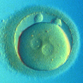
Embryo Biopsy
The first step in the PGD protocol is embryo biopsy. Embryo biopsy is the process of removing a single cell from a preimplantation human embryo. By analyzing the biopsied cell we can draw conclusions as to the genetic status of the whole embryo. If the cell removed is a male cell, we can be 100% sure that the embryo will eventually develop into a boy, if the same cell also has an extra chromosome 21, we conclude that such embryo will develop into a boy with Down's Syndrome.
Embryo biopsy can be performed on Day 3, Day 4, or Day 5 after fertilization.


Day 3 Biopsy
For the embryo biopsy an embryologist uses one of three methods to break through the zona pellucida (a glycoprotein layer surrounding the embryo, a.k.a. "zona"):
- Partial zona dissection (PZD) is done mechanically using a glass needle. This is the quickest and safest method of performing biopsy. However, this is also the most technically challenging method.
- Acidified solution is used to dissolve a hole in the zona pellucida.
- A Laser is used to burn a slit in the zona.
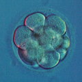
Once the zona is "opened" an individual blastomere is taken from the embryo using a biopsy pipette which is blunt, flame-polished, and has an inner diameter of about 30-50μm for the safe removal of one blastomere. Some embryologists do not feel comfortable using a biopsy pipette, they either "flush out" or "squeeze out" one or a few blastomeres through the opening in the zona.
Day 4 Biopsy
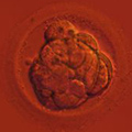
Performed in exactly the same way as Day 3 biopsy but the cell loss to the embryo is reduced by the fact that only one out of 16-30 cells is biopsied instead of one out of 6-12.
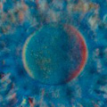
Day 5 Biopsy
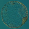
By Day 5 the embryo reaches the blastocyst stage. Biopsy on Day 5 is done with a laser to slice away a cluster of 2 to 10 or more cells. The cells biopsied on day 5 are taken from the trophectoderm (TE), which is the outer layer of the blastocyst. Following the biopsy the blastocyst is usually frozen, but sometimes it is cultured until Day 6 or even Day 7 waiting for the results of genetic analysis.
Embryo Biopsy on Day 3 vs. Day 4 vs. Day 5
At ViaGene we strongly recommend embryo biopsy to be performed on Day 3 with Embryo Transfer (ET) on Day 4 (even Day 3, for our local IVF Centers). There is no need to unnecessarily prolong embryo stay in culture, unless a lot of embryos are available for transfer and an extra selection for the "toughest" embryos is possible. It should be noted, though, that being the "toughest" in vitro does not necessarily translate into the most viable. More importantly, being unable to reach the blastocyst stage in vitro does not mean the same for in vivo conditions. Embryos which arrest by Day 5 in the culture media may have survived, if transferred by Day 4. This is especially crucial for the patients of the advanced maternal age (AMA, women over the age of 36) who may only have 1 or 2 embryos available for transfer by Day 4, and potentially none by Day 5.
(Although it might not be relevant to humans, it should be noted that in the Research Laboratories and in animal husbandry preimplantation embryos are transferred back into the oviducts or uteri of their mothers as soon as technically possible. The best results in transgenic mice project was achieved when zygotes were transferred into the ampulla region of the oviducts immediately after transgenic DNA injection into the zygote's pronucleus.)
It has been argued that Day 5 biopsy avoids the problem of embryo mosaicism by taking 10 or more cells for analysis. However, averaging all biopsied TE cells by WGA for Microarray analysis is not the best way to detect mosaicism. Furthermore, being mostly "sisters" and "granddaughter" blastomeres of a single blastomere, the biopsy of 5-10 TE cells provides us, even if analyzed individually, with genetics of the progeny of a single blastomere of the 8-cell embryo. Numerous attempts to show the effects of ooplasm polarization have shown us, if anything, that cells do not migrate at the preimplantation stage.
At ViaGene, when we do 24-chromosome aneuploidy testing for IVF Centers concerned with embryo mosaicism or the myth of self-correction, we offer Day 4 biopsy of two cells from the opposite sides of the morula. This is less damaging than taking 5-10 cells one day later and also gives us a better chance of detecting mosaicism than any other PGD technique.
Does biopsy damage the embryo?
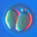
Biopsy itself, if performed by a professional, poses no risk to the embryo. However, one cell has to be removed and we should never forget that Day 3 embryos have only 8 cells. One blastomere out of 8 may seem like a lot, but this does not mean that later in development an embryo would miss one-eighth of its size, or some vital organ. The preimplantation human embryo is able to compensate for one missing cell. This is a benefit of being among the species, which use the so-called regulative type of development. In a human embryo, the fate of the blastomere is not preprogrammed by the egg. Instead, the function of the cell is determined and readjusted according to it's the position in the developing embryo.
It must be stressed that the benefit of selecting out genetically abnormal embryos must outweigh the negative effect of a single cell removal. It has been proven that unless each step of the PGD procedure is done by professionals, the results may be less than beneficial, and sometimes even damaging to the outcome of the IVF cycle. This is why it is still important to choose a reliable PGD laboratory and a qualified embryologist to perform the biopsy procedure.
Analyzing the Collected Samples
After biopsy, the removed cells must be analyzed using one of the available PGD methods. An in-depth discussion about currently the Currently Available PGD / PGS Technologies is available on the next page.