PGD Terminology
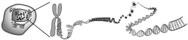
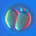
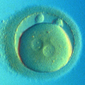
Normal Embryo: Humans have 46 chromosomes: 22 pairs of autosomes, and two sex chromosomes, X+X or X+Y. Any chromosome combination other than 46,XX (Normal Female) or 46,XY (Normal Male) is considered ABNORMAL.
Monosomy: A monosomy is an absence of one chromosome. A monosomy of any autosome is lethal in humans, only a few babies with autosomal monosomy have survived beyond birth, they all had monosomy of chromosome 21.
Trisomy: A trisomy is a presence of one extra chromosome. The most common among newborn is a trisomy of chromosome 21, leading to Down's Syndrome. According to the latest published National Vital Statistics report, out of 2,748,302 births, 1,298 had Down's Syndrome and 1,093 had other chromosomal anomalies. Among these "other chromosomal anomalies" the most common is an abnormal number of sex chromosomes. Per each 100,000 recognized pregnancies, around 1,400 abort due to an abnormal number of sex chromosomes, 100-200 boys are born with Kleinfelter syndrome (instead of XY they have XXY, XXXY, XXYY, or even XXXXY sets of sex chromosomes) and about 50 girls are born with Turner syndrome (instead of XX they have X, XXX, or XXXX).
Other common trisomies include trisomy 18 (Edward's Syndrome) and 13 (Patau's Syndrome). Frequencies of either syndrome range from 1 in 2,000 to 1 in 15,000. The only other chromosomes noticeably affecting the outcome of established pregnancies are chromosomes 16 (trisomy 16 is found in 1,229 among 15,000 spontaneous abortions) and 22 (trisomy 22 is found in 424 out of 15,000 spontaneous abortions). Aneuploidy for any other chromosome is lethal at the very first stages of embryo development, before a pregnancy can even be established.
Haploid: Haploid embryos have only one set of 23 chromosomes. They originate from parthenogenetically activated oocytes, sometimes from prematurely dividing zygotes.
Triploid, Tetraploid, or Polyploid embryos are those having full extra sets of all 23 chromosomes. These embryos originate from an oocyte fertilized by two spermatozoa, by diploid spermatozoon, or from an oocyte which failed to extrude the second polar body. Polyploidization also occurs during embryo cleavage, however, at the blastocyst stage it is a normal step in trophectoderm formation.
Chaotic Cleavage means that during mitosis, when the zygote and, subsequently, blastomeres divide into two daughter cells, the chromosomes segregate between two sister blastomeres randomly or chaotically, causing multiple monosomys and trisomys. Most of these embryos are also morphologically abnormal and very few of them progress beyond the cleavage stage. Such embryos are usually marked as having "Complex Abnormalities."
Embryo Mosaicism: Mosaicism is a result of mitotic error and as such arises during embryo cleavage, when one of the blastomeres divides into two genetically unequal daughter blastomeres. This may lead to an embryo having both normal and abnormal cells, i.e., mosaic embryo. Cells with autosomal monosomy may be selected out during further embryo development; however, if their population rises above some critical level, the embryo will die before or shortly after implantation. If an embryo is suspected of having genetically normal cells and cells with autosomal monosomy, such embryos should be avoided during embryo transfer. Since mosaic embryos with monosomy of chromosome 21 may result in a live birth, such mosaic embryos are marked as abnormal.
Embryo with genetically normal cells and cells with autosomal trisomy may develop into an abnormal mosaic baby. Some newborns with Down's, Edward's, and Patau's Syndrome are actually mosaics. Mosaic embryos with trisomies should never be considered for transfer.
Multinucleate/Anucleate Blastomere: Some embryos may be revealed as abnormal even prior to genetic testing, during embryo biopsy or after blastomere fixation. If a single blastomere has more than one nucleus, it is called a multinucleate blastomere. Even if each nucleus (separated in the PGD Report by square brackets) is genetically normal, or if they add to a normal set of chromosomes, the corresponding embryo may be genetically abnormal. Multinucleation indicates some gross abnormalities in the timing between cell division (cytokinesis) and nuclear division (karyokinesis). If cleavage results in one blastomere retaining both nuclei, then its 'sister blastomere' will have none. Absence of a nucleus (anucleate blastomere) may be similar in its origins to multinucleation, but it may also be considered as an extreme example of embryo fragmentation. Although anucleate blastomeres cannot give any indications as to the embryo's genetic background, it should be noted that their presence lowers embryo viability. If multiple morphologically normal blastomeres were analyzed, and none of them had a nucleus, such an embryo may be considered not viable due to gross errors in embryo cleavage.