Scientific Publications
• Transgenic Mice with Integrated Sequences of RSV Provirus DNA
Citation: Solomko A.P., Morozova L.M., Vapina I.N., Evsikov S.V. and Chashchina L.I. (1988) Transgenic mice with integrated sequences of RSV provirus DNA. Genome 30(suppl.):104.
Abstract: The transgenic mice containing recombinant pATV-8 plasmid, produced by insertion of the provirus DNA of RSV into pBR322 were obtained. The type of the Iransgene DNA inheritance cannot be explained by the insertion of its copies into one chromosomal site. In certain transgenic mice lines the phenotypic disorders were observed.
Full Text: Download Original Article in PDF
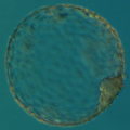
• The Role of the Nucleocytoplasmic Ratio in Development Regulation of the Early Mouse Embryo
Citation: Evsikov S.V., Morozova L.M. and Solomko A.P. (1990) The role of the nucleocytoplasmic ratio in development regulation of the early mouse embryo. Development 109:323-328.
Abstract: The hypothesis suggesting that the blastocoele is able to form only at a definite nucleocytoplasmic ratio was tested. We compared the development of preimplantation mouse embryos under different conditions. The results demonstrated that the start of cavitation is not dependent on the number of cell divisions. Thus, a definite nucleocytoplasmic ratio is not required for blastocoele formation to start. Our studies on embryos with microsurgically altered cytoplasm content provided evidence for the following biological clock mechanism: a change in the cell program of morphogenesis needs definite concentration of the products of a previous genetic program.
Keywords: nucleocytoplasmic ratio, blastocoele formation, biological clock, microsurgery, mouse embryo.
Full Text: Download Original Article in PDF
• Preconception Genetic Diagnosis of Cystic Fibrosis
Citation: Strom C.M., Verlinsky Y., Milayeva S., Evsikov S., Cieslak J., Lifchez A., Valle J., Moise J., Ginsberg N. and Applebaum M. (1990) Preconseption genetic diagnosis of cystic fibrosis. Lancet 336:306-307.
Abstract: Preconception genetic diagnosis by polar body removal has been developed for couples at high risk for conceiving genetically abnormal fetuses. The procedure involves retrieval of oocytes under ultrasound guidance, removal of the first polar body before fertilisation, and genetic analysis of the polar body using the polymerase chain reaction (PCR). In women heterozygous for a genetic disease, the polar body will, in the absence of crossing-over, be homozygous for the gene not contained in the oocyte from which it was removed. If crossing over has occurred, the polar body will be heterozygous, and it will not be possible to determine the genotype of the oocyte in this way. Pregnancies have been established following blastomere biopsy, demonstrating the feasibility of preimplantation genetic analysis.
Full Text: Download Original Article in PDF
• Fertilization of Human Oocytes by Sperm Microinjection and Electrofusion
Citation: Evsikov S.V., Cieslak J. and Verlinsky Y. (1990) Fertilization of human oocytes by sperm microinjection and electrofusion. J. In Vitro Fert. Embryo Transf. 7:190.
• Techniques for Micromanipulation and Biopsy of Human Gametes and Preembryos
Citation: Verlinsky Y., Cieslak J. and Evsikov S. (1991) Techniques for micromanipulation and biopsy of human gametes and preembryos. In: Preimplantation Genetics (ed. by Y.Verlinsky, A.Kuliev) New York, Plenum Press, pp.273-278
• Preconception and Preimplantation Diagnosis for Cystic Fibrosis
Citation: Verlinsky Y., Rechitsky S., Evsikov S., White M., Cieslak J., Lifchez A., Valle J., Moise J. and Strom C. (1992) Preconception and preimplantation diagnosis for cystic fibrosis. Prenatal Diagnosis 12:103-110.
Abstract: Preimplantation diagnosis provides couples at high genetic risk the possibility of avoiding genetic disease without the need for prenatal diagnosis and selective abortion of the affected pregnancy. Following extensive background work on the reliability of genetic diagnosis in a single cell, we offered on a research basis preimplantation diagnosis to five couples at risk for offspring with the delta-F508 mutation (the major mutation causing cystic fibrosis). There was no detrimental effect from polar body removal on either fertilization or preimplantation development. Genetic analysis, undertaken in 22 polar bodies and 15 corresponding blastomeres, identified 21 embryos of which ten were transferred.
Keywords: Preimplantation diagnosis Polar body removal Cystic fibrosis Embryo biopsy
Full Text: Download Original Article in PDF
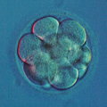
• Role of the Spatial Zygote Cytoplasm Organization in Cell Differentiation at the First Stages of Mouse Development
Citation: Evsikov S.V. and Solomko A.P. (1993) Role of the spatial zygote cytoplasm organization in cell differentiation at the first stages of mouse development. Doklady Akademii Nauk (Russia) 332:254-256.
• Role of Ooplasmic Segregation in Mammalian Development
Citation: Evsikov S.V., Morozova L.M. and Solomko A.P. (1994) Role of ooplasmic segregation in mammalian development. Roux's Arch. Dev. Biol. 203:199-204.
Abstract: Abstract. A new micromanipulation technique permitted the scrambling of the zygote cytoplasm. Such interference had no effect on preimplantation development, and when zygotes with scrambled cytoplasm were transfered to the pseudopregnant females, normal and fertile mice were born. This demonstrates that no morphogenetic factors are prelocalized in the egg cytoplasm. Cleavage characteristics of mouse embryos provide the evidence that zygote cytoplasm does not define any determinate type of cleavage. We conclude that the mechanism of ooplasmic segregation is not used in the mouse (and presumably mammalian) development. It is suggested that the turning point in the evolution of mammalian embryogenesis was the transition to the intrauterine development, that started the process leading among other changes, to the loss of the ooplasmic morphogenetic determinants.
Keywords: Cell differentiation, Cytoplasm, Micromanipulation, Mouse embryo, Evolution
Full Text: Download Original Article in PDF
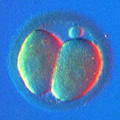
• Preimplantation Development of Manipulated Mouse Zygotes Fused with Second Polar Bodies
Citation: Evsikov S.V. and Evsikov A.V. (1994) Preimplantation development of manipulated mouse zygotes fused with the second polar bodies: a cytogenetic study. Int. J. Dev. Biol. 38:725-730.
Abstract: Immediately after fertilization one chromatid of each maternally-derived chromosome is extruded into the second polar body (2PB). We tested the ability of these "extra" chromosomes to support preimplantation development. Micromanipulation and electrofusion techniques were used to fuse 2PBs with diploid, haploid, or enucleated mouse zygotes. Androgenetic haploids, intact embryos, and digynic triploids served as the controls for the reconstructed embryos. Androgenetic haploid zygotes developed to the blastocyst stage only when fused with the 2PBs. This result demonstrates that even when extruded into the 2PB, chromosomes retain their ability to support normal preimplantation development. However, 2PB fusion with diploid zygote impaired preimplantation development. Normal development of experimentally produced digynic triploids (zygotes with one extra maternal pronucleus) indicated that developmental arrests. caused by the 2PB fusion. were not the resu It of triploidy or micromanipulation procedures. Cytogenetic studies showed that developmental failures of the reconstructed embryos were caused by premature chromosome condensation of the polar body chromosomes. This result indicates that 2PB must be removed from the zygotes' perivitelline space during animal cloning experiments. In addition, we showed that 2PB fusion with enucleated zygote is a reliable method for 2PB karyotyping and may be used in the studies of mammalian meiosis.
Keywords: Mouse embryo, polar body, premature chromosome condensation, triploid, haploid.
Full Text: Download Original Article in PDF
• Visualisation and Cytogenetic Analysis of Second Polar Body Chromosomes Following its Fusion with a One-Cell Mouse Embryo
Citation: Verlinsky Y., Dozortsev D. and Evsikov S. (1994) Visualisation and cytogenetic analysis of second polar body chromosomes following its fusion with a one-cell mouse embryo. J.Assisted Reprod.Dev. 11:123-131.
Abstract: This study was designed to visualize the second polar body (2PB) chromosomes using its electrofusion with a one-cell-stage mouse embryo to approach preconception diagnosis of chromosomal disorders. Results: Eighty to 90% hybridization efficiency has been achieved by electrofusion of 2PB with mouse zygotes. 2PB chromosomes were visualized in 40-50% of hybrids. Sixty-five percent of 2PB chromosomes were visualized when fused with the cytoplast obtained microsurgically by removing pronuclei from a one-cell embryo. As much as 33-43% of these resulting metaphases appeared to contain chromosomal aberrations. The follow-up of the development of the reconstructed one cell-stage hybrids in vitro revealed a significant decrease in their viability. The hybrid embryos resulting from 2PB electrofusion with enucleated zygotes did not develop beyond the two-cell stage. Conclusion: Electrofusion is an efficient approach for hybridization of 2PB with a one-cell mouse embryo and may be useful for visualization and cytogenetic analysis of 2PB chromosomes. The visualization rate of 2PB chromosomes is higher if 2PB is fused with enucleated zygotes. However, the method induces over 30% of chromosomal aberrations and may lead to a significant decrease in the viability of the resulting one-cell embryos.
Keywords: second polar body (2PB), 2PB chromosomes, preimplantation diagnosis, electrofusion.
Full Text: Download Original Article in PDF
• Investigation of the Biological Clock Mechanism Operating at the Preimplantation Stage of Mouse Development
Citation: Evsikov S.V. and Solomko A.P. (1994) Investigation of the biological clock mechanism operating at the preimplantation stage of mouse development. Biol. Reprod. 50(Suppl.1):127.
• Embryo Viability and the Timing of Blastocoel Formation Depend on the Nucleocytoplasmic Ratio of the Reconstructed Mouse Embryos
Citation: Evsikov S.V. (1995) Embryo viability and the timing of blastocoel formation depend on the nucleocytoplasmic ratio of the reconstructed mouse embryos. Proc. of the Congress of the European Developmental Biology Organisation, Toulouse, France, July 9-13, 1995, p.137.
• A Study of the First and Second Polar Bodies in Mouse Oogenesis
Citation: Evsikov A.V. and Evsikov S.V. (1995) A study of the first and second polar bodies in mouse oogenesis. Ontogenez 26:196-200 (In Russian).
Abstract: The possibility of using a polar body for biopsy of human oocytes and early embryos has recently been shown. Genetic analysis of polar bodies can also provide additional information about the mechanisms underlying oocyte maturation. The first polar body is extremely unstable: in mouse oocytes, it disintegrates within several hours. Thus, the possibilities for its analysis are limited. We obtained karyoplasts of mouse eggs that contained the metaphase II spindle. By using them as a model for the first polar body, we studied the causes of its rapid disintegration. The rates of disintegration of the karyoplasts treated with various inhibitors of the cytoskeleton indicate that disintegration of the first polar body may be due to interaction between the actin cytoskeleton, chromatin, and the plasma membrane. The second polar body is found in mouse embryos until the blastocyst stage. Fusion of the second polar body with the enucleated zygote allowed analysis of its chromosomes.
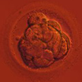
• Mechanisms of Cell Number Regulation in the Per-Implantation Mouse Blastocyst
Citation: Evsikov S.V., Vagyna I.N. and Solomko A.P. (1996) Mechanisms of cell number regulation in the per-implantation mouse blastocyst. J.Exp.Zool. 276:201-208.
Abstract: Low viability of manipulated or in vitro cultured embryos is caused primarily by the reduced cell number in the implanting blastocysts. In order to investigate the effect of implantation delay on embryo viability and cell number, mouse blastocysts were transferred into oviducts of day 0 pseudopregnant females. This type of transfer improved embryo survival rates, indicating that embryos retarded by in vitro culture restored their viability during 3 days of delayed implantation. Our results showed that even in the cases when the initial cell count was as low as 28.2 0.7 cells per blastocyst (vs. 60.5 f 1.4 cells in the control blastocysts, developed in vivo), implantation delay increased this number to 107.2 2 3.5 cells (control blastocysts had at this stage on average 111.0 3.7 cells). Half-blastocysts, developed from the single blastomeres of the 2-cell embryos or from experimentally produced tetraploids, had around 50 cells after 3 days of implantation delay. This indicates that the start of blastocyst dormancy is triggered during the eighth cell cycle and independent of the absolute cell number or the number of cytokineses. Implantation delayed blastocysts, developed from the half-embryos with the doubled volume of cytoplasm, had on average 70.5 * 2.4 cells, suggesting that embryo fall into quiescence is also dependent upon the attainment of a definite nucleo-cytoplasmic ratio. We conclude that blastocyst readiness for implantation is determined by two factors: number of cell cycles and nucleo-cytoplasmic ratio.
Full Text: Download Original Article in PDF
• Factors Affecting Readiness of Mouse Blastocysts for Implantation
Citation: Vagyna I.N., Evsikov S.V., and Solomko A.P. (1997) Factors Affecting Readiness of Mouse Blastocysts for Implantation. Biopolimery i Kletka 13:161-167.
Abstract: Mechanisms of cell number regulation in mouse blastocysts have been investigated using embryo transfers into the oviducts of pseudopregnant Day 0 females. It was shown that the retarded after in vitro culture embryos are overtaking in their cell number the in vivo developed embryos during the period of implantation delay. Studies on cell numbers of blastocysts developed from one blastomere of 2-cell embryo, and experimentally produced tetraploids, demonsrate that blastocysts are entering the pre-implantation diapause during the 8-th cell cycle. This process does not depend upon absolute cell number or number of cell divisions. Blastocysts, which developed from the half-embryos with doubled cytoplasm content, had on average 70.5±2.4 cells. This observation proves that blastocyst entering to the diapause depend upon reaching of definite nucleo-cytoptasmic ratio. It seems that the readiness of blastocysts for implantation depends on two factors, number of cell cycles and nucleo-cytoplasmic ratio.
Full Text: Download Original Article in PDF
• Prepregnancy Genetic Testing for Age-Related Aneuploidies by Polar Body Analysis
Citation: Verlinsky Y., Cieslak J., Ivakhnenko V., Evsikov S., Wolf G., White M., Lifchez A., Kaplan B., Moise J., Valle J., Ginsberg N., Strom C., and Kuliev A. (1997) Genetic Testing 1:231-235.
Abstract: Current practice for prevention of chromosomal aneuploidies involves prenatal screening and termination of pregnancy, a procedure that is not universally acceptable. We introduced prepregnancy genetic testing by sampling and fluorescence in situ hybridization (FISH) analysis of the first and second polar body (PB), to avoid fertilization and transfer of embryos resulting from aneuploid oocytes. In 395 in vitro fertilization (IVF) patients of advanced maternal age, the first and second PBs were removed following their extrusion from oocytes and studied by FISH, using probes specific for chromosomes 13, 18, and 21, to detect and avoid the transfer of oocytes with common aneuploidies. Overall, 3,651 oocytes obtained from 598 IVF cycles were available for FISH analysis, with 2,952 showing interpretable FISH results (80.9%). The analysis revealed 1,271 (43.1%) oocytes with aneuploidy, which were excluded from transfer and subjected to follow-up FISH analysis to confirm PB diagnosis in the cleavage or blastocyst stage embryos. Only embryos originating from 1,681 aneuploidy-free oocytes were transferred back to patients, resulting in 119 pregnancies overall, from which 78 healthy children have already been born, 35 were spontaneously aborted, and 16 are ongoing, after confirming PB diagnosis by prenatal diagnosis. The results demonstrate that PB-based preimplantation diagnosis may be used for prepregnancy screening in women with age-related risk for common aneuploidies.
Full Text: Download Original Article in PDF
• Preimplantation Diagnosis of Thalassemias
Citation: Kuliev A., Rechitsky S., Verlinsky O., Ivakhnenko V., Evsikov S., Wolf G., Angastiniotis M., Georghiou D., Kukharenko V., Strom C. and Verlinsky Y. (1998) Preimplantation diagnosis of thalassemias. J. Assist. Reprod. Genet. 15:219-225
Abstract: Purpose: Preimplantation genetic diagnosis (PGD) is an important option for couples at risk of having children with B-globin mutations to avoid selective abortions of affected fetuses following prenatal diagnosis. Methods: We performed PGD for thalassemia in 12 clinical cycles (IVS1-110, and IVS-745 mutations) using biopsy of the first and second polar bodies (PBs) extruded from oocytes during maturation and fertilization, coupled with nested polymerase chain reaction analysis and restriction digestion. Results: A total of 118 oocytes was obtained, of which 78 had results for both the first and the second PBs. This resulted in the selection and transfer of 30 unaffected embryos (2.5 embryos per cycle). To avoid a possible misdiagnosis due to allele dropout (ADO), we have also introduced simultaneous detection of two highly polymorphic linked markers, a short tandem repeat immediately at the 5' end of the globin gene and HUMTH01 which is a syntenic short tandem repeat. The application of multiplex polymerase chain reaction of the B-globin gene and linked polymorphic markers enabled detection of ADO in five first PBs, thus avoiding the transfer of potentially affected embryos resulting from their corresponding oocytes. Conclusions: Confirmation studies of the embryos resulting from the oocytes predicted to contain an affected gene confirmed the diagnosis in 98% of the cases, thus demonstrating the accuracy and reliability ofPB PGD of thalassemia mutations. The application of PB analysis in six patients resulted in two ongoing pregnancies with a thalassemia-free fetus already confirmed in both of them by prenatal diagnosis.
Keywords: preimplantation genetic diagnosis, multiplex PCR, thalassemia, polymorphic markers, B-globin gene, allele dropout.
Full Text: Download Original Article in PDF
• Preimplantation Diagnosis of Common Aneuploidies by the First- and Second-Polar Body FISH Analysis
Citation: Verlinsky Y., Cieslak J., Ivakhnenko V., Evsikov S., Wolf G., White M., Lifchez A., Kaplan B., Moise J., Valle J., Ginsberg N., Strom C. and Kuliev A. (1998) Preimplantation diagnosis of common aneuploidies by the first- and second-polar body FISH analysis. J. Assist. Reprod. Genet. 15:285-289.
Abstract: Purpose: A low pregnancy rate in in vitro fertilization (IVF) patients of advanced maternal age may be caused by aneuploidies originating from non disjunction in the first or second meiotic divisions. We introduced genetic testing of oocytes by sampling and fluorescent in situ hybridization (FISH) analysis of the first and second polar bodies, to avoid fertilization and transfer of aneuploid oocytes in IVF patients of advanced maternal age. Methods: Three hundred and sixty-three IVF patients 34 years and older participated in the study. Using micromanipulation procedures, the first and second polar bodies were removed following their extrusion from the oocytes and studied by FISH, using probes specific for chromosomes 13, 18, and 21 to detect oocytes with common aneuploidies. Results: Of a total of 538 IVF cycles, 3250 oocytes were available for FISH analysis, with conclusive FISH results in 2742 oocytes (84.3%). As many as 1102 (40%) of oocytes were predicted to be aneuploid and not transferred. Of 1640 embryos predicted to be normal, 1145 were transferred in 467 treatment cycles, resulting in 107 pregnancies (23%), from which 67 healthy children have been born, 32 pregnancies spontaneously aborted, and 15pregnancies are ongoing after being confirmed normal by prenatal diagnosis. Conclusions: Preimplantation diagnosis by first- and second- polar body FISH analysis allows us to avoid the agerelated risk of common aneuploidies in IVF patients of advanced maternal age.
Keywords: preimplantation diagnosis; first polar body; second polar body; fluorescent in situ hybridization (FISH) chromosome 13-, 18-, and 21 specific probes; chromatid errors; common aneuploidies.
Full Text: Download Original Article in PDF
• Mosaicism in the Inner Cell Mass of Human Blastocysts
Citation: Evsikov S. and Verlinsky Y. (1998) Mosaicism in the inner cell mass of human blastocysts. Hum. Reprod. 13:3151-3155.
Abstract: Although mosaicism was shown to be a normal feature in cleaving embryos, its consequences for the late preimplantation stages are unknown.We performed blastocyst immunosurgery, followed by fluorescent in-situ hybridization (FISH), to determine the number of cells and degree of mosaicism in the inner cell mass (ICM) of human blastocysts. Of 47 ICM samples analysed, 20 had aneuploid cells, and two also had a few tetraploid cells. The average degree of aneuploidy in the ICM was similar to the overall blastocyst mosaicism, suggesting that there is probably no selection for euploid ICM. The lower degree of blastocyst mosaicism, compared with the cleavage-stage embryos, may be due to a mechanism of selection against the embryos with high frequency of mosaicism, leading to elimination of these embryos prior to blastocyst formation.
Keywords: aneuploidy, FISH, human blastocyst, inner cell mass, mosaicism.
Full Text: Download Original Article in PDF
• Karyotyping of Human Oocytes by Chromosomal Analysis of Second Polar Bodies
Citation: Verlinsky Y. and Evsikov S. (1999) Karyotyping of human oocytes by chromosomal analysis of the second polar bodies. Mol. Hum. Reprod. 5:89-95.
Abstract: This paper describes a method for obtaining metaphase chromosomes from human second polar bodies. The second polar body nucleus was injected into the cytoplasm of an enucleated oocyte, which is activated shortly after injection. When the polar body nucleus is transformed into a haploid pronucleus, treatment with okadaic acid was used to induce premature chromosome condensation. A total of 25 analysable chromosome plates were obtained from 38 polar bodies karyotyped using this technique. Whole chromosome painting was used to detect second polar bodies (and respectively, oocytes) with unbalanced translocations. In combination with the first polar body analysis, this technique may be useful in preimplantation genetic diagnosis for patients carrying maternal translocations.
Keywords: metaphase chromosomes, micromanipulations, oocyte activation, polar body, whole chromosome painting.
Full Text: Download Original Article in PDF
• Visualization of Chromosomes in Single Human Blastomeres
Citation: Evsikov S. and Verlinsky Y. (1999) Visualization of chromosomes in single human blastomeres. J. Assist. Reprod. Genet. 16:133-137.
Abstract: FISH analysis of the first polar bodies has been used to detect chromosomal translocations in human oocytes. However, this technique is limited for maternally derived translocations and since, at present, there is no reliable method for visualizing blastomere chromosomes, paternally derived translocations cannot be detected at the preimplantation stage. Nuclear transplantations have been extensively used for the study of nucleocytoplasmic interactions, cell differentiation, and animal and embryo cloning. It is usually expected that heterologous nuclei, if transferred into enucleated eggs and zygotes, would undergo remodeling in accordance with the developmental program of the ooplasm. In this study, we investigated the possibility of applying nuclear transplantation techniques for karyotyping a single blastomere of preimplantation human embryo. Our expectations were based on the fact that individual blastomeres, when fused with ooplasts, will be involved in the cell cycle, characteristic for recipient ooplasm and thus, the timing of mitosis could be manipulated and predicted.
Full Text: Download Original Article in PDF
• Prevention of Age-Related Aneuploidies by Polar Body Testing of Oocytes
Citation: Verlinsky Y., Cieslak J., Ivakhnenko V., Evsikov S., Wolf G., White M., Lifchez A., Kaplan B., Moise J., Valle J., Ginsberg N., Strom C. and Kuliev A. (1999) Prevention of age-related aneuploidies by polar body testing of oocytes. J. Assist. Reprod. Genet. 16:165-169.
Abstract: Purpose: We previously demonstrated that aneuploidy-free oocytes may be preselected by testing the first and second polar bodies removed from oocytes following their maturation and fertilization. The present paper describes the results of the application of the method in 659 in vitro fertilization cycles from patients of advanced maternal age. Methods: Using micromanipulation techniques, 3943 oocytes were tested by polar body sampling and fluorescent in situ hybridization analysis using specific probes for chromosomes 13, 18, and 21. Results: Fluorescent in situ hybridization results were available for 3217 (81.6%) of 3943 oocytes studied, of which 1388 (43.1%) had aneuploidies; 35.7% of the aneuploidies were of first meiotic division origin, and 26.1% of second meiotic division origin. Most errors in the first meiotic division were represented by chromatid malsegregation. The transfer of embryos deriving from 1558 of 1829 aneuploidyfree oocytes in 614 treatment cycles resulted in 131 clinical pregnancies and 88 healthy children born after confirmation of the polar body diagnosis. Conclusions: Polar body testing of oocytes provides an accurate and reliable approach for prevention of age-related aneuploidies in in vitro fertilization patients of advanced maternal age.
Keywords: preimpiantation diagnosis, chromosome 13, 18, and 21 aneuploidies, FISH, first and second polar bodies, IVF.
Full Text: Download Original Article in PDF
• Birth of Healthy Children After Preimplantation Diagnosis of Thalassemias
Citation: Kuliev A., Rechitsky S., Verlinsky O., Ivakhnenko V., Cieslak J., Evsikov S., Wolf G., Angastiniotis M., Kalakoutis G., Strom C. and Verlinsky Y. (1999) Birth of healthy children after preimplantation diagnosis of thalassemias. J. Assist. Reprod. Genet. 16:207-211.
Abstract: Background: Preimplantation genetic diagnosis (PGD) allows couples at risk of having children with thalassemia to ensure the healthy outcome of their pregnancy. Methods: Seventeen PGD clinical cycles were initiated for Cypriot couples at risk of having children with different thalassemia mutations, including IVS1-110, IVSI-6, and IVS 11-745. Unaffected embryos for transfer were selected by testing oocytes, using first and second polar body (PB) removal and nested polymerase chain reaction analysis followed by restriction digestion. Results: Unaffected embryos were selected in 16 of 17 PGD cycles. Of 166 oocytes studied from these cycles, 110 were analyzed by sequential analysis of both the first and the second PB, resulting in preselection and transfer of 45 unaffected embryos. This resulted in seven pregnancies and in the birth of five healthy thalassemia-free children. The embryos predicted to have inherited the affected allele were not transferred. Analysis of these embryos confirmed the PB diagnosis. Conclusions: Sequential first and second PB testing of oocytes is reliable for PGD of thalassemia and is a feasible alternative to prenatal diagnosis in high-risk populations.
Keywords: preimplantation genetic diagnosis, thalassemia, multiplex nested PCR, allele dropout.
Full Text: Download Original Article in PDF
• A Simplified and Efficient Method for Obtaining Metaphase Chromosomes from Individual Human Blastomeres
Citation: Verlinsky Y. and Evsikov S. (1999) A simplified and efficient method for obtaining metaphase chromosomes from individual human blastomeres. Fertil. Steril. 72:1127-1133.
Abstract: Objective: To develop a reliable and cost-effective technique for karyotyping single human blastomeres for preimplantation diagnosis of chromosomal translocations. Design: Controlled laboratory study. Setting: Preimplantation genetic diagnosis and IVF program, Reproductive Genetics Institute/IVF Illinois, Chicago, Illinois. Patient(s): Patients undergoing IVF and preimplantation genetic diagnosis. Intervention(s): Individual human blastomeres were fused with enucleated or intact mouse zygotes. After blastomere– cytoplast fusion, heterokaryons were fixed at metaphase of the first cleavage division or treated with okadaic acid to induce premature chromosome condensation. Main Outcome Measure(s): Percentage of analyzable metaphase plates and ease and reliability of the procedure. Result(s): The effectiveness of the proposed technique with blastomeres from day 3 diploid embryos was 91%. Sixty-three metaphases were obtained from 69 blastomeres; 3 blastomeres had not fused, 1 heterokaryon had no chromatin (an anucleated cytoplasmic bleb was biopsied and fused), and 2 heterokaryons cleaved before they were fixed. Conclusion(s): Human blastomere fusion with an intact mouse zygote is an efficient and technically undemanding method for obtaining metaphase chromosome plates from individual human blastomeres for preimplantation testing for chromosomal translocations and aneuploidy.
Keywords: Chromosomal translocations, preimplantation genetic diagnosis, karyotyping, visualization of chromosomes, whole chromosome painting.
Full Text: Download Original Article in PDF
• Effect of Chromosomal Translocations on the Development of Preimplantation Human Embryos In Vitro
Citation: Evsikov S., Cieslak J. and Verlinsky Y (2000) Effect of chromosomal translocations on the development of preimplantation human embryos in vitro. Fertil. Steril. 74:672-677.
Abstract: Objective: To determine the reliability of a new technique for single human blastomere karyotyping during clinical cases for preimplantation genetic diagnosis of translocations. Design: Controlled clinical study. Setting: Preimplantation genetic diagnosis and IVF program. Patient(s): Nineteen preimplantation genetic diagnosis cases with 11 types of translocations (10 reciprocal and one Robertsonian) involving chromosomes 1, 5, 7, 8, 9, 11, 12, 13, 14, 15, 16, 18, 20, 21, and 22. Intervention(s): Blastomere biopsy followed by blastomere nucleus conversion into metaphase chromosomes. Fluorescent in situ hybridization (whole chromosome painting) was used for the detection of chromosomally unbalanced preimplantation human embryos. Main Outcome Measure(s): Percentage of informative metaphase plates and effect of unbalanced translocations on preimplantation embryo development. Result(s): Informative metaphases were obtained for 84% of the blastomeres. Analysis of preimplantation development of the resulting embryos showed that an unbalanced chromosomal complement does not affect embryo ability to reach the blastocyst stage in vitro. Conclusion(s): For the translocations tested, there is no evident selection against chromosomally unbalanced embryos at the preimplantation stage of embryo development.
Keywords: Blastocyst development, blastomere biopsy, chromosome translocation, FISH, metaphase chromosomes, preimplantation genetic diagnosis.
Full Text: Download Original Article in PDF
• High Frequency of Meiosis II Aneuploidies in IVF Patients of Advanced Maternal Age.
Citation: Verlinsky Y., Cieslak J., Ivakhnenko V., Evsikov S., Strom C., Kuliev A. (2000) Fertility and Sterility, October 2000, S33.
Abstract: It is well established that most chromosomal abnormalities originate from female meiosis, contributing considerably to pregnancy failures particularly in women of advanced maternal age. Based on DNA polymorphism studies in children with chromosomal syndromes and their parents, it was suggested that these abnormalities are mainly of meiosis I origin, although no direct observations of the outcomes of the first or second meiotic divisions have been available so far. The results present the first demonstration of the high frequency of aneuploidies originating from the second meiotic division, suggesting the clinical significance of detection and avoidance from transfer of the embryos resulting from the oocytes with meiosis II errors in IVF patients of advanced maternal age.
Full Text: Download Original Article in PDF
• Text Book: An Atlas of Preimplantation Genetic Diagnosis
Citation: S.Evsikov is one of the major contributors to "An Atlas of Preimplantation Genetic Diagnosis" by Y.Verlinsky and A.Kuliev, Parthenon Publishing, London, 2000.
Abstract: This color-illustrated atlas and clinical textbook by two respected authorities details the procedures for diagnosing genetic disease in gametes prior to fertilization and in embryos fertilized in vitro prior to uteral implantation. With over 200 full-color images, the book sets out the current methods used in oocyte and preembryo sampling along with the latest techniques in DNA and cytogenetic analysis in preimplantation development. Includes bibliographic references and index. Authors Verlinsky and Kuliev are well-known experts on this subject with two previous books to their credit.
Full Text: Buy on Amazon
• Chromosomal Abnormalities in the First and Second Polar Body
Citation: Verlinsky Y., Cieslak J., Ivakhnenko V., Evsikov S., Wolf G., White M., Lifchez A., Kaplan B., Moise J., Valle J., Ginsberg N., Strom C. and Kuliev A. (2001) Chromosomal abnormalities in the first and second polar body. Mol. Cell. Endocrinol. 183(Suppl.1):S47-49.
Abstract: Aneuploidy free oocytes may be pre-selected by testing the first and second polar bodies removed from oocytes following their maturation and fertilization. We present here our experience on the application of the method in IVF cycles from patients of advanced maternal age. Overall, 5590 oocytes were obtained from 917 cycles and tested by polar body sampling and fluorescent in situ hybridization (FISH) analysis using specific probes for chromosomes 13, 16, 18, 21 and 22. FISH results were available in 4599 (82.2%) of 5590 oocytes studied, from which 2077(45.2%) were with aneuploidies. Thirty six point one percent of aneuploidies were of the first meiotic origin, and 29.3% of the second meiotic origin. Most errors in the first meiotic division were represented by chromatid errors. The transfer of embryos deriving from 2014 of 2520 aneuploidy free oocytes in 821 treatment cycles resulted in 182 (22.2%) clinical pregnancies and 140 healthy children born after confirmation of the polar body diagnosis. Polar body testing of oocytes provides an approach for pre-selection of aneuploidy free embryos, improving pregnancy rate in IVF patents of advanced maternal age.
Keywords: preimplantation diagnosis, Chromosome 13, 16, 18, 21 and 22 aneuploidies, FISH, first and second polar body, IVF patients of advanced maternal age
Full Text: Download Original Article in PDF
• Nuclear Transfer for Full Karyotyping and Preimplantation Diagnosis for Translocations
Citation: Verlinsky Y., Cieslak J., Evsikov S., Galat V. and Kuliev A. (2002) Nuclear transfer for full karyotyping and preimplantation diagnosis for translocations. J. Reprod. Biomed. Online 5:300-305.
Abstract: Preimplantation genetic diagnosis (PGD) for chromosomal disorders is currently perfomed by interphase fluorescence insitu hybridization (FISH) analysis. This technique is known to have limitations for detecting some translocations and complete karyotyping, so visualization of chromosomes in single cells is required to improve the PGD accuracy for chromosomal disorders. To achieve this, single blastomeres were fused with enucleated or intact mouse zygotes, followed by fixing the resulting heterokaryons at the metaphase of the first cleavage division, or treating them with okadaic acid to induce premature chromosome condensation. This method alowed a significant improvement in the accuracy of testing both maternally- and paternally-derived translocations. In all, 437 blastomeres were tested, achieving full karyotyping in as many as 383 (88%), making it possible to pre-select only normal embryos or those with balanced chromosomal complements for transfer. Overall, PGD for translocations was applied in 94 clinical cycles, resulting in 66 transfers and 20(30.3%) clinical pregnancies, with healthy deliveriess of 15 children. Fifty-two of these cycles were performed using a nuclear transfer (conversion) technique, which resulted in 38 transfers of balanced or normal embryos, demonstrating that the techinique is accurate and reliable for karyotyping single blastomeres for PGD of translocations.
Keywords: blastomere nuclear conversion, chromosomal translocations, FISH, mouse oocytes, nuclear transfer, PGD
Full Text: Download Original Article in PDF
• The Genetic Control of Mammalian Early Embryogenesis
Citation: Vagyna, I.N., Evsikov, S.V., and Solomko, A.P. (2003) The Genetic Control of Mammalian Early Embryogenesis. Biopolimery i Kletka 19:3-10.
Abstract: The review focuses on the problems of genetic control of mammalian early development. The modern views on the processes of pre-implantation development and implantation of eutherian embryos are discussed.
Full Text: Download Original Article in PDF